Copyright ©2026 IMAGINE ECHO. All Rights Reserved

Full-Service Home Diagnostic Solutions


.png)
Early screening supports heart health by helping identify potential concerns sooner—empowering you and your healthcare provider to make informed care decisions.

Enjoy the comfort and privacy of home-based screenings. No need to travel – we bring professional healthcare to your doorstep.

Trusted by healthcare professionals in the Bay Area, our services support proactive heart health screening.

Each screening is conducted with the utmost care and attention, ensuring a personalized experience tailored to your needs.
Physicians can partner with us for value-based care.

Convenient, affordable cardiac & sleep care delivered to you — with onsite echocardiogram and EKG services, home-delivered diagnostics, fast 24–48 hour results, and hospital-quality testing at competive rates.

Comprehensive ultrasound examination of your heart to assess its structure and function.

Non-invasive test to monitor your heart's electrical activity and identify any irregularities.
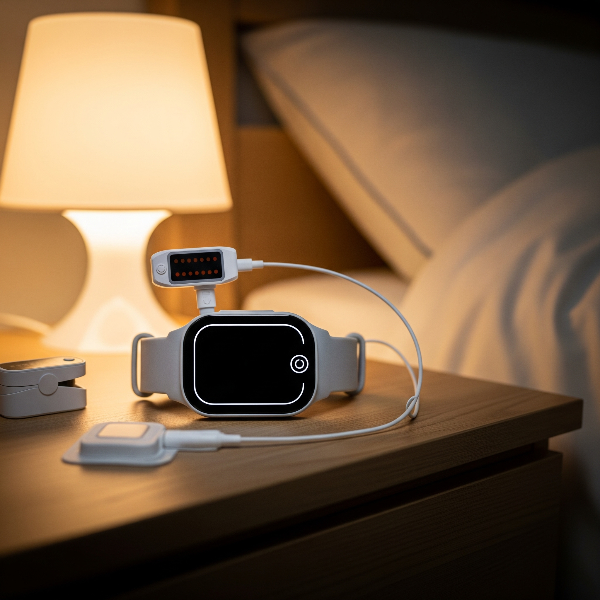
Most advanced home sleep test clinically proven to detect obstructive sleep apnea and central sleep apnea.
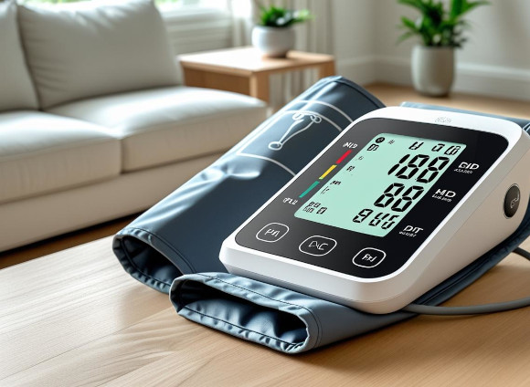
24-hour ambulatory blood pressure monitor tracks day & night readings, providing a detailed profile to manage hypertension with expert review.
Heart Home Screening is a comprehensive evaluation of your heart health conducted in the comfort of your home. It includes various non-invasive tests to assess the risk of heart disease and other cardiovascular conditions.
Each screening session typically takes about 45 minutes, depending on the specific assessments included. Our team ensures a thorough and efficient process.
No, our screening process is entirely non-invasive and pain-free. We use advanced equipment to monitor your heart health without any discomfort.
Our service offers the unique advantage of convenience and comfort by bringing the screening to your home. We provide personalized attention and use state-of-the-art equipment, ensuring quality care without the need for you to visit a hospital or clinic.
Our screenings are conducted by certified and experienced healthcare professionals who specialize in cardiovascular health. They are trained to provide a comfortable and informative experience.
No special preparation is needed. We recommend wearing comfortable clothing and avoiding heavy meals or vigorous exercise immediately before the screening.
We use the latest in medical technology, including advanced Ultrasound, ECG machines, blood pressure monitors, and other diagnostic tools to ensure accurate and reliable results.
Absolutely. We provide a detailed report of your screening results, which you can keep for your records or share with your healthcare provider.
Reports are provided within 24 hours. All exams read by US based board certified cardiologist within 24 hours